Our Technology
The Intraoral Camera: Seeing What We See
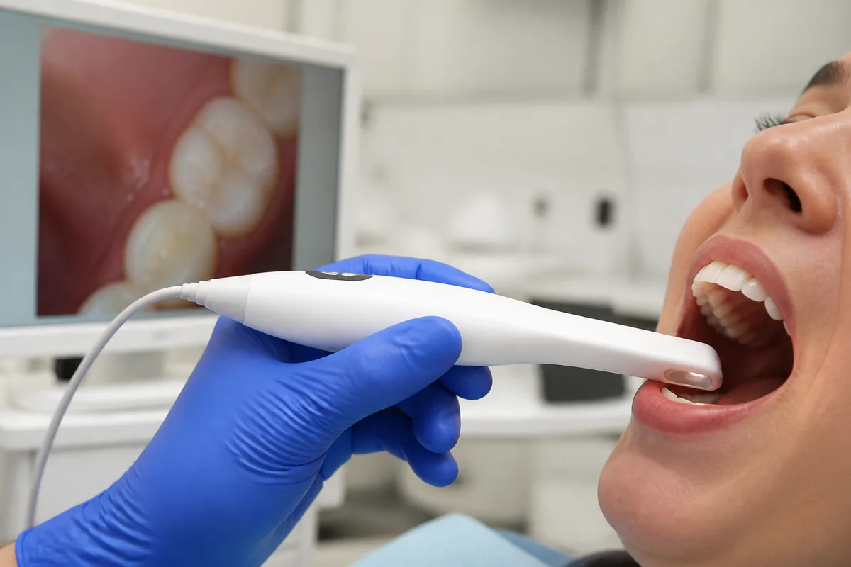
An intraoral camera is a slim wand, about the size of a large pen, with a lens and a ring of light at the tip. It captures high-resolution still images and live video of individual teeth and displays them on the monitor beside the chair, magnified many times over.
It is the least technically complicated piece of equipment in a modern operatory and arguably the one that changes the patient experience most, because it closes the gap between what the dentist can see and what you can.
How it works
The camera has a wide depth of field and its own light source, so it produces a sharp, evenly lit image of a surface a few centimeters away in a dark, wet, curved space. Magnification is typically in the range of twenty to sixty times, which turns a hairline crack into something obvious.
Images are captured with a button or a foot pedal and stored directly in your chart, which means they become part of your record. A photograph of a tooth taken this year can be placed beside one from three years ago, and a change that would be impossible to describe in words is immediately visible.
The tip is covered with a disposable sheath for each patient, and the body is disinfected between uses. It is entirely painless and involves no radiation.
What it shows
Cracks and craze lines in enamel, which are extremely hard to see with the naked eye and are often the explanation for a tooth that hurts on biting. Fractured cusps and chipped edges. Wear facets from grinding, which are flat polished areas that tell a story about how your jaw moves at night.
Failing restorations: the dark line at the margin of an old filling, the gap where a crown meets the tooth, the stained edge where leakage has begun. Recurrent decay beneath a restoration when it reaches the surface. Broken or worn-out fillings that feel fine and look fine from a distance.
Soft tissue too: inflamed gum margins, recession, a lesion or ulcer that needs following up, plaque and tartar accumulation in the areas you are missing when you brush. Seeing exactly where the plaque is on your own teeth is a more effective home-care lesson than any general advice about technique.
Why seeing it matters
Dentistry asks people to consent to treatment for problems they cannot see, often in a tooth that does not hurt. That is a genuine ask, and skepticism about it is reasonable.
An image removes most of that difficulty. When you can see the crack running across your own molar, a recommendation to crown it stops being an assertion and becomes an observation you can evaluate for yourself. Equally, when we say a small area is stable and worth watching rather than treating, an image lets us show you the same thing next year and demonstrate that it has not changed.
It is also useful for second opinions, for insurance claims where a photograph supports the diagnosis, and for communicating with a specialist about a specific finding before you get there.
What it cannot do
The camera photographs surfaces. It cannot see between teeth where they touch, it cannot see inside a tooth, and it cannot see bone. Decay starting between two teeth, infection at a root tip and bone loss from gum disease are all diagnosed on X-rays, not photographs.
So it complements imaging rather than replacing it, and a complete picture usually needs both. It also does not replace the clinical examination: some findings are detected by touch and by how a tooth responds to testing rather than by appearance.
If you would like to see what is going on in your own mouth rather than hear about it, ask at your next visit and we will put it on the screen. Call Cedar Creek Dentistry in Portland.
Questions about your own care? The team at The Best Dentist In Portland, Oregon is happy to help — call us at (503) 646-1811.
This article is for general information only and is not medical advice.