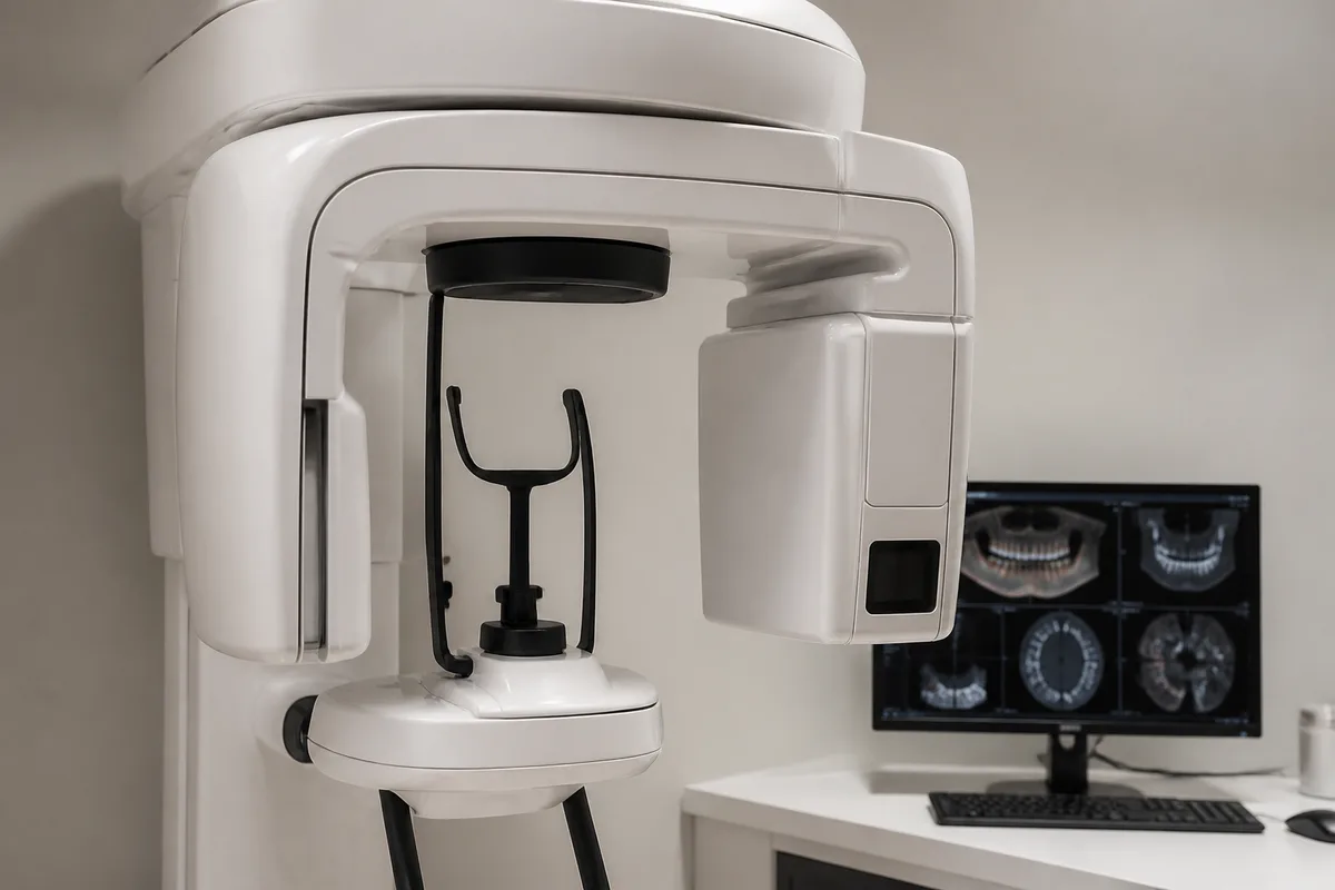
Our Technology
Panoramic X-Rays: The Whole Picture in One Image
A panoramic X-ray, often called a Panorex after an early brand, is the single wide image that shows both jaws end to end, all the teeth, the sinuses above and the jaw joints at each side, on one picture.
It is taken with the machine rotating around your head while you stand or sit still, takes under twenty seconds, and requires nothing inside your mouth, which makes it the easiest dental image to take for anyone with a strong gag reflex.
How the image is made
The X-ray source and the sensor are mounted on opposite ends of an arm that rotates around your head. Because the source and sensor move in a synchronized arc, the machine keeps a curved layer of tissue, the shape of the dental arch, in sharp focus while everything nearer and further blurs out.
That curved focal trough is why the resulting image looks like the jaws have been unrolled flat. It is a reconstruction rather than a straightforward photograph, which explains both its strengths and its distortions.
Positioning matters more than on any other dental image. Your chin rests on a support, you bite on a positioning stick, and you are asked to keep your tongue against the roof of your mouth and stay completely still. Small positioning errors produce characteristic distortions, which is why the technician is fussy about it.
What it shows
Wisdom teeth, including impacted ones still fully inside the bone, their angle, and their relationship to the nerve canal in the lower jaw. This is the most common reason a panoramic image is taken.
Developing teeth in children and adolescents: how many there are, whether any are missing or extra, and whether any are erupting off course. An impacted upper canine caught at age nine is a very different problem from one found at fifteen.
Also: cysts and tumors in the jaws, which can grow silently to considerable size; fractures; the jaw joints and gross asymmetry between the two sides; bone level in broad terms; retained roots from old extractions; the sinus floor above the upper back teeth, which matters for implant planning; and occasionally calcification in the carotid artery in the neck, an incidental finding worth mentioning to a physician.
What it does not show well
A panoramic image is not a substitute for bitewings. Its resolution is lower, and the teeth at the front overlap on the image, so early decay between teeth is regularly missed on it. If you need cavity detection, you need bitewings.
Fine detail generally is limited. Small areas of infection at a root tip, subtle bone changes and the precise margin of a restoration are better assessed on an intraoral image.
It is also two-dimensional and geometrically distorted, with magnification that varies across the image, so it cannot be measured reliably for implant planning. That is what a cone beam scan is for.
Dose and when it is appropriate
A digital panoramic image delivers a low dose, broadly comparable to a few days of natural background radiation and considerably less than a full mouth series of intraoral films. Digital sensors reduced it substantially compared with film systems.
It is typically taken for a new comprehensive examination, for wisdom tooth assessment in the late teens, during orthodontic evaluation, before extractions or implant planning, when investigating jaw pain or swelling, and then updated at fairly long intervals rather than routinely.
As with all dental imaging, the right frequency depends on what question is being asked rather than on a calendar. If you have had a panoramic image taken elsewhere recently, tell us and we will request it rather than repeat it. Call Cedar Creek Dentistry in Portland.
Questions about your own care? The team at The Best Dentist In Portland, Oregon is happy to help — call us at (503) 646-1811.
This article is for general information only and is not medical advice.