Our Technology
Digital X-Rays: Less Radiation, Better Diagnosis
Digital radiography replaced film in dentistry over the past two decades, and the change was not cosmetic. A digital sensor needs a fraction of the radiation film required to produce an image, the image appears on screen in about a second, and it can be adjusted, magnified and compared against previous years without ever leaving the room.
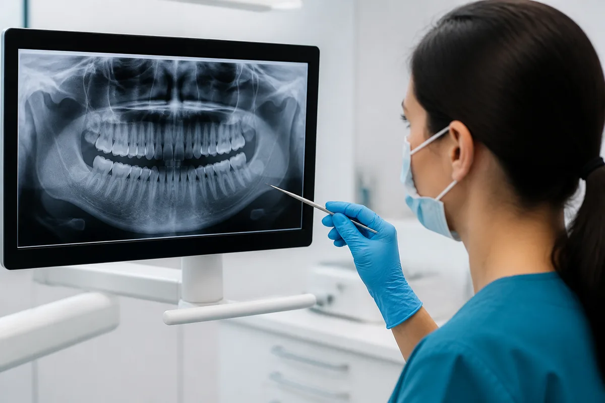
The practical result is that we see more, sooner, with less exposure, and you can see the same image we are looking at.
How they work
A small electronic sensor replaces the film packet. It converts the X-rays that pass through your teeth directly into a digital signal, which is sent to the computer and rendered immediately. There is no chemical processing, no darkroom, and no waiting.
Because the sensor is far more sensitive to radiation than film emulsion, a much shorter exposure produces a usable image. Reported reductions compared with traditional film commonly range from fifty to eighty percent depending on the film speed being compared against.
Once on screen, the image can be enlarged, its contrast and brightness adjusted, and specific areas examined closely. A shadow that is ambiguous at normal size can often be resolved by adjusting the display, which sometimes avoids retaking an image entirely.
Putting the dose in perspective
Everyone is exposed to background radiation continuously, from the ground, from building materials, from food and from cosmic rays. In the United States that background is roughly three millisieverts a year.
A set of four digital bitewing X-rays represents a very small fraction of a single day's background exposure. A full mouth series is larger but still a small fraction of the annual background. For comparison, a commercial flight across the country delivers a measurable dose from cosmic radiation.
The profession still works to the principle of keeping exposure as low as reasonably achievable. That means a lead apron with a thyroid collar where appropriate, rectangular collimation that narrows the beam to the shape of the sensor, and, most importantly, only taking images when there is a diagnostic reason to. Tell us if you are or may be pregnant; dental X-rays with proper shielding are considered safe in pregnancy, but non-urgent imaging is usually deferred anyway.
What they show that looking cannot
Decay between teeth where the surfaces touch is the most common finding. That contact area is invisible to the eye and cannot be probed, and a cavity can be well established there before any symptom appears. Bitewing images are specifically designed to show it.
Bone level around the teeth is the objective record of periodontal disease, and comparing this year to five years ago answers questions that no single image can. Infection at the root tip appears as a dark area in the bone, often in a tooth that does not hurt. Cysts, impacted teeth, retained roots, resorption, and abnormalities in the jaw are all radiographic findings.
Images also show what is under existing work: decay beneath the margin of a crown or an old filling, a poorly sealed root canal, an overhanging restoration catching floss. None of that is visible from above.
How often, and the practical advantages
There is no universal interval. Frequency should follow your individual risk: someone with a history of frequent decay, active periodontal disease, dry mouth or extensive restorative work reasonably has bitewings more often than someone with no restorations, healthy gums and no decay in a decade. A full mouth series or a panoramic image is usually taken for a new comprehensive examination and updated at much longer intervals.
Digital images are also easy to share. They transfer instantly to a specialist, to your insurance company for a pre-authorization, or to you by email if you are moving. Bringing recent images from a previous dentist can save you a set.
And having the image on a large screen beside you changes the conversation. When we say there is decay between two teeth, you can see the dark area yourself instead of taking it on trust. If you have questions about imaging or want your records sent somewhere, call Cedar Creek Dentistry in Portland.
Questions about your own care? The team at The Best Dentist In Portland, Oregon is happy to help — call us at (503) 646-1811.
This article is for general information only and is not medical advice.